Accueil » WHAT IS TEM?
About Transmission Electron Microscopy (TEM)
What is the principle of TEM ?
Electron microscopy is a powerful tool for the investigation of materials using a very high magnification (X10,000) and high-resolution (0,1nm). The materials are analysed to their elemental composition the results are then compared to already known reference materials. This enables the lab analyst to achieve accurate and precise identification of samples to determine if they are, in-fact asbestos. TEM also features the ability to identify non-commercial asbestos types such as tremolite, actinolite and anthophyllite which cannot be identified by Polarised Light Microscopy (PLM).
The concept of the TEM is to produce electron at the top of the microscope within the electron gun by heating a tungsten filament which will send, through a vacuum column, electrons through the sample. The sample is disposed at the middle of the vacuum column by a specific sample holder.
Comparing to SEM where electrons will reflect on the sample, here electrons will transmit the sample. All electrons will then hit the fluorescent screen enable the image recording system to produce the image you are looking for.

Main characteristics
The source of illumination
Compared to Light Microscopy (LM), the Transmission Electron Microscopy (TEM) is built at the opposite. The source of illumination is at the top of the microscope and the screen at the bottom The source of illumination comes from the electron gun which by heating a tungsten or crystal filament generates electrons transmitting the sample all way through the microscope within a high speed. Electrons are accelerated in the gun section within a voltage between 80kV and 200 kV. The thicker is your sample, the higher voltage you need to penetrate the sample.
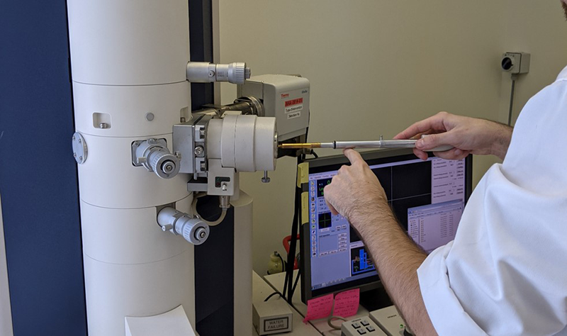
The Sample Holder
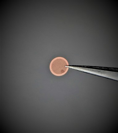
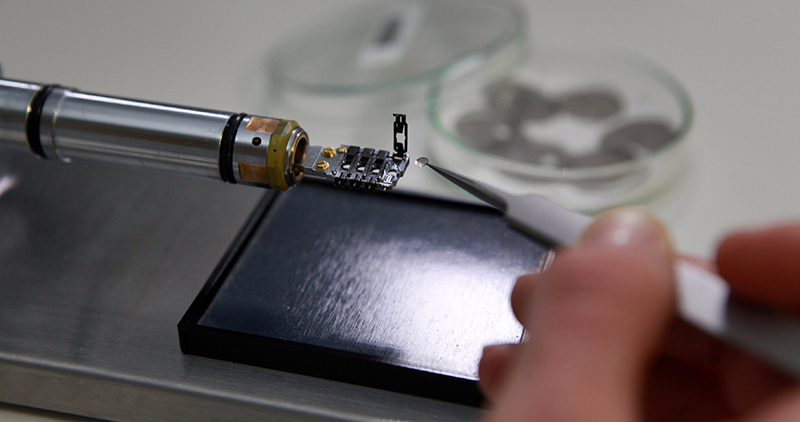
The sample/specimen holder allows the insertion of samples into the vacuum with a minimal loss on the column thanks to an O-ring that separates the atmospheric pressure from the vacuum. A vacuum pump evacuates the air on the pre-chamber before inserting the sample in the column. The sample preparation is put on a metal grid about 3 mm of diameter. We usually use a copper grid. The metal grid gets various mesh size ranging from a few to 100 microns. The TEM stage allows the movement of the sample in the XY plan to locate the region of interest.
The Apertures
We distinguish 3 kind of apertures within the TEM. One Condenser aperture where you chose the size of the beam (small spot size of the beam, loose intensity but increase the accuracy); one Objective aperture which you can select out and filter out the electrons that deflected in the sample to increase contrast; and one Intermediary aperture which enable for the electron diffraction to select the region of interest on the sample.
The Vacuum System
The TEM is equipped with various vacuum systems enabling the machine to create vacuum in the column essential for electron microscopy. The vacuum is needed to allow the voltage isolation between the electromagnetic lens working as a cathode and the electron without generating an arc. The vacuum is also needed to reduce collision frequency of electrons with gas atoms.
What is the principle of TEM ?
The analyst makes his decision depending on 3 final criteria :
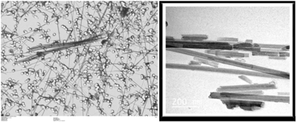
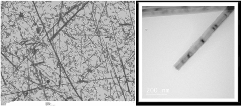
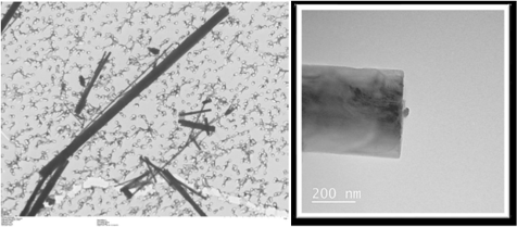
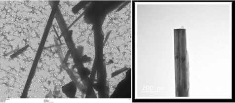
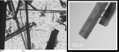
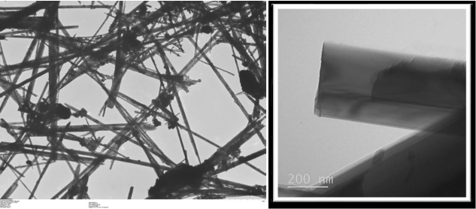
Morphology
The analyst looks deep into the sample and investigates all fibrous shapes that matches with the definition of an asbestiform.
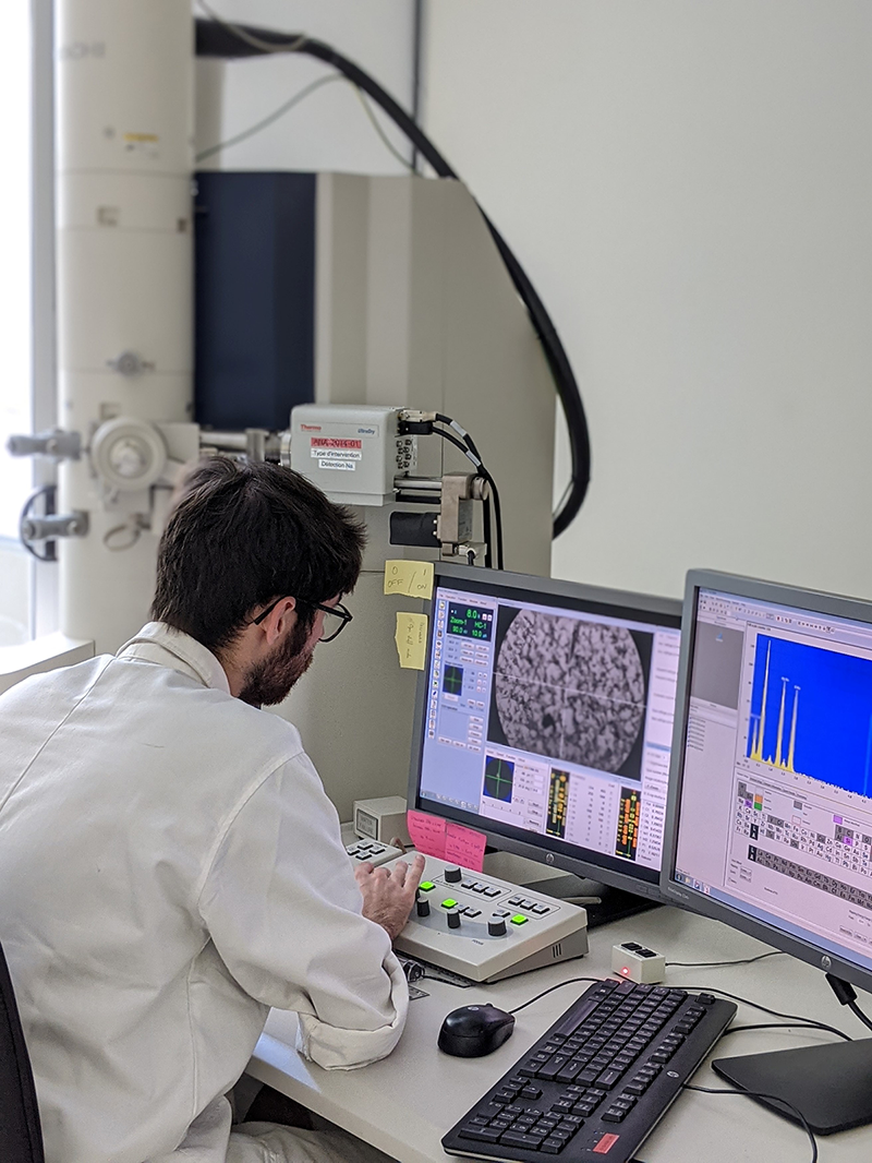
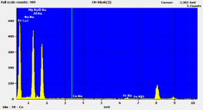
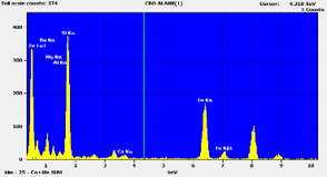
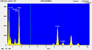
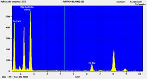
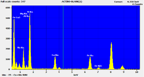
Chemistry
An Energy-Dispersive X-ray Spectroscopy (EDS) is used for element characterisation. Comparison of the specimen’s spectrum with the spectra of asbestos known compositions produces qualitative results.
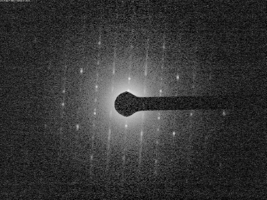
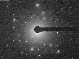
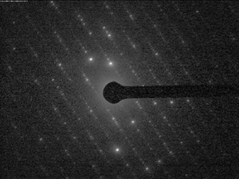
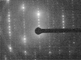
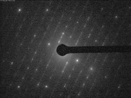
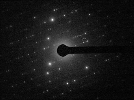
Crystal structure
Defined with convergent beam electron diffraction technique called SAED (Selected Area Electron Diffraction). The atomic arrangement pattern of the structure observed is then qualitatively compared to reference patterns of asbestos to identify the mineral.
3 criteria that allows unequivocal identification of asbestos including the thinest fibrils of chrysotile (0.02um in width) undetectable by PLM, PCM, and Low-resolution SEM.